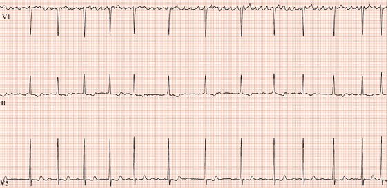
Multi-electrode radiofrequency catheters have several electrodes, each of which can deliver radiofrequency energy. If all the dots are not connected, afib could re-enter the heart in the unablated space (gap). To make lesions, electrophysiologists ablate one spot after another, similar to drawing a line by making dots one after the other. Single electrode radiofrequency catheters emit radiofrequency energy from a single point at the catheter tip. Here are the different types of catheters available today for atrial fibrillation catheter ablations: It’s believed that different types of catheters could have higher success rates or fewer complications. Most atrial fibrillation ablations today use single point radiofrequency energy catheters, which have a success rate-freedom from atrial fibrillation-of about 70%. The design and functionality of catheters is constantly evolving. Other energy sources, such as microwave and high intensity focused ultrasound (HIFU), have been studied but these energy sources aren’t widely used in catheter ablation. In addition, laser energy can be adjusted when ablating tissues of varying thickness, with higher energy applied to thicker tissue and lower energy applied to thinner structures in the heart. Unlike other energy sources, contact force is not a factor in whether a laser lesion is transmural. With laser energy, the catheter doesn’t need to be in direct contact with tissue, which could mean it will have fewer complications than radiofrequency energy. It may be a safer heat-based energy than unipolar radiofrequency energy. Laser energy is light waves that are converted to heat to ablate and create scar tissue. Cryo energy can be used in single point catheters or balloon catheters. Animal studies have shown cryo energy to have a much lower incidence of thrombus (clot) formation than radiofrequency energy, which suggests that cryothermy could lower the risk of a stroke. In addition, cryo energy may have fewer major complications. It may be easier to make lesions using cryothermy since the cold temperatures cause tissue to stick to the catheter. Traditional catheters use unipolar radiofrequency energy, which simply means that the radiofrequency energy is transmitted from a single point at the catheter’s tip.Ĭryo energy, is intense cold that is used to form ice crystals within the tissue, causing the cells to die and creating scar tissue. In general, high temperatures are needed to make sure that all layers of tissue are ablated. It reliably achieves transmurality and is the most widely used energy source in catheter ablations. Radiofrequency (RF) energy is radio waves that are converted to heat to ablate and create scar tissue. When the abnormal pathway is destroyed, the heart’s electrical impulses can only travel through the normal conduction pathways, and the arrhythmia is eliminated. Radiofrequency energy is a form of electrical energy that causes the tissue at the tip of the catheter to heat up, permanently damaging (ablating) that small area of tissue. Most commonly used energy is called Radiofrequency (RF). At this point, an ablation is performed.Ī special catheter will be used to direct energy to the abnormal pathway in the heart.

Then, in a process called mapping, a catheter (small, flexible tube) is manipulated until it locates the abnormal pathway that is causing the tachycardia. The EP Study helps identify the specific type and location of the abnormal heart rhythm. If you haven’t already had an EP Study, the two procedures are frequently performed one after the other. Often, this treatment permanently resolves the arrhythmia by destroying the abnormal pathway that causes it.Ī cardiac ablation is very similar to an Electrophysiology (EP) Study. Your physician may choose to treat your arrhythmia with a non-surgical procedure called cardiac ablation. These arrhythmias can be treated, but not cured, with medications. These abnormal pathways can make the electrical impulses that control your heart rate travel in a circular pattern within the heart, causing a tachycardia. SVT may occur when an extra electrical pathway exists in or between some of the structures of the heart. If the tachycardia originates in one of the upper chambers of the heart, it”s called a supraventricular (above the ventricles) tachycardia (SVT).

When the heart beats too rapidly, the arrhythmia is called a tachycardia. Ordinarily, your heart beats at a regular, steady pace called a normal sinus rhythm that is regulated electrically by the sinus node.


 0 kommentar(er)
